
Bioanalytical Protein-Protein Detection with the P4SPR
SPR platform based on ionic liquids for the detection of a breast cancer biomarker in cell lysates
The detection of biomarkers in real biological samples poses a real challenge in medical diagnostics. Samples may be blood, serum, plasma, cell lysate, urine, and saliva; these biofluids contain numerous cellular components that are amenable to high biofouling on any sensor surfaces. Cell lysates, in particular, have a higher lipid content than serum [1]. Thus, any biosensing surfaces that were previously thought to be suitable to detect biomarkers in serum while minimizing non-specific adsorption, such as hydrophilic PEG-based surfaces, may not work as well for cell lysates.
An alternative to hydrophilic surfaces is to use ionic liquids. Surface chemistries that are hydrophobic and positively charged have been observed to reduce non-specific adsorption from cell lysate samples [1]. Thus, the research group led by Prof. Masson at Université de Montréal screened a number of ionic liquids and other surface chemistries for their level of non-specific adsorption in the presence of cell lysates [1]. This blog briefly summarizes their findings of how an ionic liquid was selected to build a surface plasmon resonance (SPR) platform to detect a breast cancer biomarker, HER2, in cell lysates. SPR is an ideal technique for point-of-care analysis due to being label-free, sensitive, and portable, and Affinité Instruments’ P4SPR embodies these sought-after key features. With the aid of a secondary detection, the HER2 biomarker was detected in undiluted cell lysates.
Evaluation of ionic liquid and other surfaces for non-specific adsorption
The surface chemistry used for biosensing must enable immobilization of the desired capture ligand and minimize biofouling. Some of the surfaces studied by Masson’s group were very efficient at reducing the non-specific adsorption, for example, dodecanethiol and 2000 Da methoxy-PEG. However, they cannot be used to immobilize capture ligands due to the lack of chemically reactive groups. Peptide monolayers that reduced non-specific adsorption from serum samples did not perform as well with cell lysate [1]. Needless to say, bare Au performed the worst in terms of non-specific adsorption level, as well as 16-mercaptohexadecanoic acid (16-MHA), which did not reduce adsorption level much more than a bare Au surface [1].
The first part of the study concluded that all the screened ionic liquids performed well and similarly in terms of reducing the level of biofouling. After a thorough study of alkyl chain length, counterions, and immobilization efficiency, [(HS)12C12(COOH)5C5im]+Br (Fig. 1) was chosen as the monolayer used on the SPR surface to detect HER2 in cell lysate [1]. The SH- group allows for self-assembly onto the gold surface while the -COOH group allows for covalent linkage to antibodies (via NH2-) through its activation by EDC/NHS* chemistry (Fig. 1). One important observation was that the level of biofouling suppression was reduced when the capture antibody was immobilized [1]. The fouling caused by SK-BR-3 breast cancer cell lysate on the anti-HER2 capture antibody-modified ionic liquid monolayer was 10-fold higher than the ionic liquid monolayer without the immobilized antibodies.
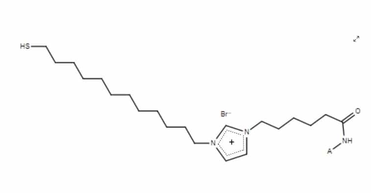
Fig. 1. Structure of ionic liquid
[(HS)12C12(COOH)5C5im]+Br when reacted with the NH2 group from an antibody. A represents an antibody.
Detection of HER2 biomarker in cell lysate
To overcome the increase in biofouling on antibody-modified ionic liquid surfaces and the low concentration of HER2 in cell lysate, the researchers implemented a secondary detection method, that is, a sandwich assay in which a different polyclonal antibody was introduced to bind to the captured biomarker [1]. The control sample used was MCF-7 cells, which contains low levels of HER2 [1]. Another MCF-7 sample was spiked with recombinant HER2 as a positive control. Finally, the third sample was the SK-BR-3 cells that should contain the HER2 biomarker.
Fig. 2 shows the schematic diagrams that represent the workflow in the case of SK-BR-3 samples. First, the anti-HER3 capture antibody-modified ionic liquid monolayer on a SPR sensor chip was exposed to SK-BR-3 cell lysates. Then, HER3 antigens were bound to the capture antibodies. The third step involved washing the SPR chip with PBS to remove cell lysate components that did not bind to the capture antibodies. Lastly, anti-HER3 detection antibodies were added, which bound specifically to the anti-HER3 antigens. Then, SPR shifts were calculated. The SPR shifts were between 7-10 times higher for the HER3-spiked MCF-7 sample and the SK-BR-3 cells compared to the MCF-7 cells only sample (control). This demonstrated that the sandwich detection using two polyclonal antibodies by SPR was able to efficiently detect HER2 biomarkers in cell lysate.
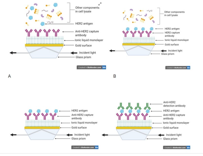
Fig. 2. A) Cell lysate containing HER2 antigens is injected into the P4SPR; B) HER2 antigens bind to anti-HER2 capture antibodies; C) Unbound cell lysate components are washed away by a PBS rinse; D) Detection of HER2 antigen is achieved by injection of anti-HER2 detection antibodies.
Conclusions
Through optimization and design, the HER2 biomarker from a crude breast cancer cell lysate was detected on a novel ionic liquid-based biosensing surface by SPR. The ionic liquid [(HS)12C12(COOH)5C5im]+Br- was used due to its ability to minimize fouling from cell lysate samples and be activated via EDC/NHS* chemistry to attach to the desired capture antibody. A secondary detection method involving a different antibody proved to be essential in observing the signal of HER2 biomarker in cell lysate. These results show promise in using ionic liquids to develop biosensing surfaces for SPR analysis for a variety of crude cell lysate samples. This is just one of many applications of the P4SPR. With research innovation in mind, Affinité Instruments can help clinical researchers develop assays with the desired surface chemistry on a SPR platform to meet global medical challenges.
*EDC= N-ethyl-N’-(3-dimethylaminopropyl)carbodiimide hydrochloride; NHS = N-hydroxysuccinimide
The Affinité Advantage
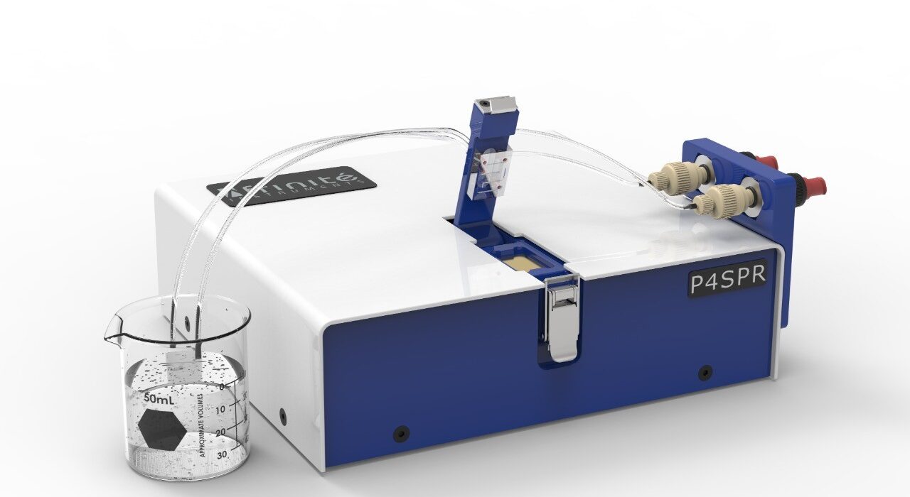
Affinité Instruments’ P4SPR™ is a very user-friendly instrument that is equipped with a gold sensing chip can be tailored to accommodate all types of surface chemistries. In addition, samples do not need much sample preparation and can be directly injected into the instrument. The P4SPR™, compared to a traditional immunoassay such as ELISA, provides fast, real-time affinity and/or kinetic data.
Simplicity – Fast training, fast results
Versatility – Pharmaceutical, biosensing, assay development applications
Economy – Affordable, accessible
Like to know more?
There is more information here: Affinité – P4SPR or give us a call on (01582) 704807
References
[1] Alexandra Aubé, Shirley Campbell, Andreea R. Schmitzer, Audrey Claing, and Jean-François Masson, Analyst, 2017, 142, 2343-2353.